Introduction
For primary breast sarcoma, wide surgical resection is a primary modality and following adjuvant radiotherapy is also known to have survival advantages. Given an adequate surgery, which requires radical resection, 5-year disease-free survival rates of breast sarcoma range from 44% to 74% [1]. Radical resection often results in considerable skin and soft tissue defect on chest wall which demands local or free flap coverage for chest wall stability and durable soft tissue coverage. Here, we introduce a case of reconstruction of chest wall defect with free DIEP flap coverage.
CASE
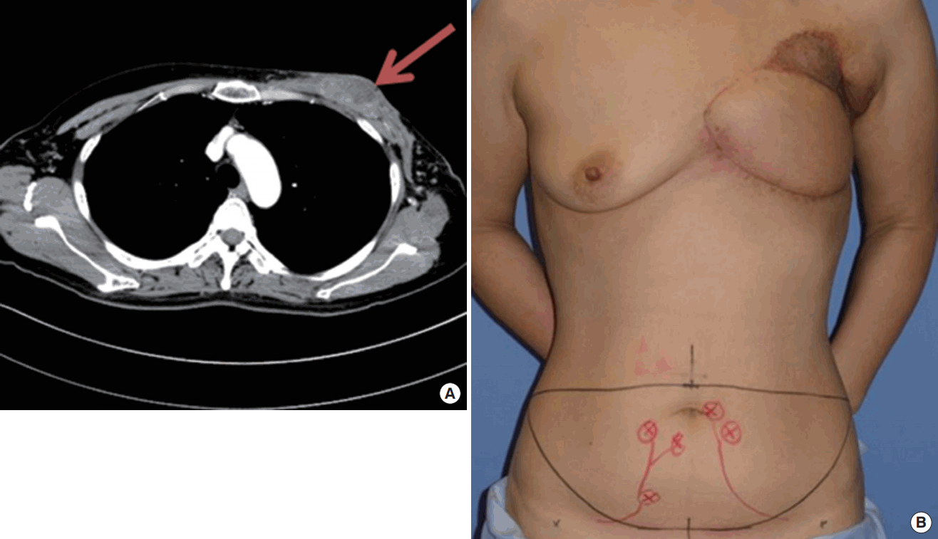
A 47-year-old female received simple mastectomy for left breast sarcoma and wound coverage with pedicled latissimus dorsi musculocutaneous flap and split thickness skin graft 5 months ago. The patient received post-operative radiation and chemotherapy but recurrent tumor mass was found on chest wall at one month follow up chest CT, which made debulking operation necessary to reduce the tumor burden (Fig. 1A).
On preoperative evaluation, complete excision of a large size defect on anterior chest wall was expected: tumors, infected and irradiated soft tissues, the underlying ribs, and sternum. Considering the substantial volume of skin and soft tissue required, and previous utility of latissimus dorsi muscle, the DIEP flap was planned for the coverage.
Preoperative computed tomography was taken to confirm the reliable blood flow of the inferior epigastric perforator vessels and identification of perforator vessels. Preoperative markings were drawn including DIEP flap of 40 cm×13 cm size and location of main perforator vessels (Fig. 1B & C). The cardiothoracic surgeon conducted en bloc chest wall resection with 2nd to 5th rib resection, upper half sternotomy and bony reconstruction with Gore-tex membrane (Fig. 2). The size of the defect was measured 25 cm×22 cm. The Internal mammary vessels and thoracoacromial vessels were exposed with intact patency and chosen for microsurgical anastomosis as recipient vessels but the thoracodorsal vessel was found compromised due to adjuvant radiation therapy. Left deep inferior epigastric artery and vein (×2) were anastomosed with left thoracoacromial artery and vein while right deep inferior epigastric artery and vein were anastomosed with internal mammary artery and vein (Fig. 3A). The defect was completely filled with the free DIEP flap as bilateral pedicle vessels comprised the whole flap with Zone I and II regions minimizing possible partial flap failure (Fig. 3B). The patient received two days of ventilator support in the intensive care unit and total hospital stay of 13 days. The pathology result revealed remnant cancer cell on the basal margin of the excised mass and owing to the durable reconstructed flap, postoperative radiation could initiate at one month post-operative period without delay. Robust flap was seen at two month after reconstruction; there was no marginal necrosis or fat necrosis and no donor site complication occurred (Fig. 3C).
Discussion
Chest wall involving breast sarcoma often requires radical resection that results in huge defect. To cover the defect and provide durable tissue, a solid flap is mandatory for the coverage. Most commonly used flaps to cover the anterior chest wall are the pectoralis major muscle flap, latissimus dorsi flap and rectus abdominis flap [2]. However, former two flaps are often inadequate for huge defects and have limited skin paddle. The rectus abdominis can be transposed on the superior epigastric pedicle or as a free flap. Free deep inferior epigastric perforator (DIEP) flap provides large volume of soft tissue with robust blood supply with limited donor-site morbidity. Yet, unilateral DIEP flap has the limitation in utilizing maximal volume of the flap; Zone IV of the flap is typically discarded due to lack of reliable perfusion. Bilateral DIEP flap provides enhanced vascular perfusion of the lower abdominal flap territory, allowing for harvest of larger volumes of tissue [3]. Enriched perfusion offers better flap survival, faster wound healing, minimal fat necrosis and marginal necrosis; thus resulting in better aesthetic outcome maintaining the initial flap design without disfig.ment. The chest wall has plenty of donor vessels that can be used for bilateral anastomoses, including thoracodorsal vessel, thoracoacromial vessel and internal mammary vessel. There have been reports of DIEP flap usage in chest wall reconstruction, but all of them were unilateral and didn’t cover the defect as huge as of the above case. (550 cm2) [4,5]. However, bilateral DIEP flap requires meticulous dissection of pedicle vessels and multiple microsurgical anastomoses. For the surgeons with extensive microsurgical experience, and when the defect requires huge volume to cover, bilateral DIEP flap would be a valuable option for anterior chest wall reconstruction as it provides substantial volume of durable tissue with minimal donor site morbidity and excellent aesthetic outcome.
Conclusion
We present the first case of free bilateral DIEP flap for the reconstruction of chest wall. When the defect is huge and donor vessels are not injured, bilateral DIEP flap would be a valuable option as it provides substantial volume of durable tissue with minimal donor site morbidity and optimal aesthetic outcome.